Mission
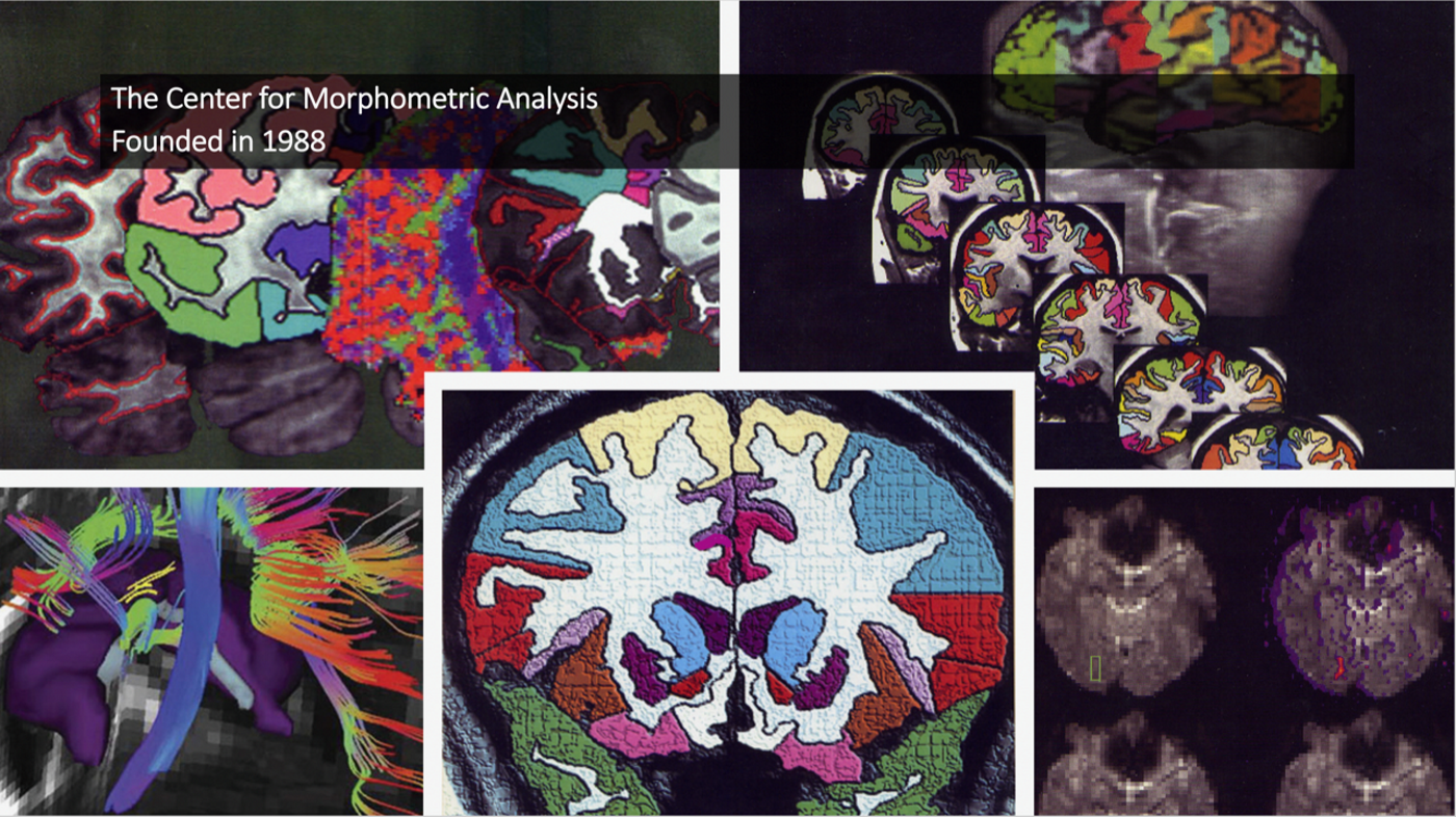
The mission of the Center for Morphometric Analysis (CMA) at Massachusetts General Hospital (MGH), Harvard Medical School, Boston, is to generate methodologies for quantitative brain anatomy inspired by developmental, comparative and clinical perspectives. The end goals of these methods are applications to basic human and ‘non-human primate’ neuroanatomy as well as to neuropsychiatric conditions to inform diagnosis, guide treatments and elucidate mechanisms of disease processes. The CMA develops targeted methods for addressing specific clinical problems using imaging technology inspired by its collaborations and informed by neurobiological knowledge and theory.
The current scientific and medical tradition of the CMA has its origins in two principal sources. The first is the Caviness-Kennedy morphometric framework, technological development and methodologies, and clinical applications of the field of magnetic resonance imaging (MRI) brain volumetrics. The second is the Geschwind-Galaburda-Pandya connectionist framework for structural and functional neuroanatomical systems in humans and non-human primates. This duality of traditions is reflected in the CMA's organizational structure and its publications in both basic neuroanatomy and clinical applications using multimodal neuroimaging since the late 1980s.
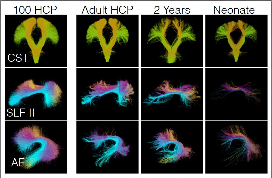
The long-term goal of our work is to create a framework of the structural ontology for the human brain to be used in the clinical domain. Therefore, classical neuroanatomy, histology and connectivity as well as all modalities of structural neuroimaging, including their validation and integration, are critically important features of our studies and the focus of our ongoing development of methodologies for basic neuroscience research and clinical applications.