The Center for Morphometric Analysis (CMA)
Departments of Psychiatry, Neurology and A. A. Martinos Center for Biomedical Imaging, Massachusetts General Hospital
History and Contributions of the Center for Morphometric Analysis
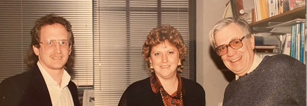
The CMA was established in the Department of Neurology at Massachusetts General Hospital in 1988. The late Dr. Verne Caviness, then Chief of Pediatric Neurology and later Interim Chief of Neurology at MGH, with colleagues Dr. David Kennedy, a nuclear physicist from M.I.T., and Dr. Pauline Filipek, a child neurologist at MGH, founded the CMA in the “non-wet bench” end of the sixth floor of the MGH Charlestown Navy Yard campus.
The CMA was among the first centers internationally to develop computer assisted programs for precise MRI volumetric quantitative imaging of the human brain. Among the notable achievements of the CMA group, it was Dr. Kennedy who developed the algorithm for MRI volumetry and also the co-registration program for the first demonstration of fMRI by a team headed by the late Dr. Jack Belliveau, a Harvard biophysicist. Dr. Nikos Makris, trained in psychiatry, stroke lesion analysis and systems neuroanatomy, pioneered a structural connectivity framework for MRI based on classical human and comparative literature on white matter connections in the brain. In addition, the CMA team led by Jim Meyer, a computer scientist and artist, developed CardViews (Cardinal Views), a neuroanatomical imaging analysis program with unique visualization and editing capabilities specifically designed for neuroanatomical structural delineation, morphometry and localization. The morphometry techniques developed by the CMA as of 1999 achieved the first complete MRI-based morphometric framework for the human brain and established a new field of science, “MRI brain volumetrics” (Caviness et al., 1999).
Moreover, the CMA disseminated this knowledge and contributed numerous brain atlases to the neuroimaging community, most notably the original human Harvard Oxford Atlas ((hHOA; see link: Harvard-Oxford cortical and subcortical structural atlases), one of the most popular cortical atlases available as part of the FMRIB Software Library (FSL) and used by neuroscientists worldwide. Critically, the CMA provided the gold standard for validation of the fully automated FreeSurfer subcortical segmentation and cortical parcellation system (Fischl et al., 2002; Fisch et al., 2004). More recently, the CMA introduced the monkey Harvard Oxford Atlas (mHOA), which shares a common homological framework with the hHOA (Rushmore et al., 2020). The hHOA and mHOA atlases embody the legacy work and knowledge reflected in the anatomical ontologies and procedures of the CMA framework in both the human and the non-human primate brain, and also provide critical neuroanatomical information for the SPL/3D Slicer brain atlas. Current efforts at the CMA aim to generate and disseminate more fine-grained, state-of-the-art, high-resolution whole-brain MR atlases of gray matter structures and their detailed structural connectivity in humans and non-human primates. The resulting atlases will be distributed to the neuroanatomical community.
Given the close relationships between the psychiatry and neurology departments at MGH, the CMA is affiliated primarily with the Department of Psychiatry and is considered an integral part of both departments within the MGH A. A. Martinos Center for Biomedical Imaging.
CMA Research Areas and Leadership, 1988 to present
The history of the CMA comprises three chronological phases as follows. From 1988 to 1999, the MRI brain volumetrics framework was developed. The period from 2000 to 2008 entailed the validation of the FreeSurfer automated MRI brain morphometric analysis by the CMA, and the development of semi-automated hybrid methods for morphometric analysis of the cerebrum, brainstem and cerebellum. Finally, from 2009 to the present the MRI comparative brain morphometric analysis framework was developed using semi-automated methodologies. Since its founding, the work and achievements of the CMA have been inspired by its many collaborators and personnel. A detailed account of these colleagues and their accomplishments within the CMA can be found at (see link: Makris & Kennedy bibliography). It is important to note that the CMA’s collaborators and personnel have helped sustain and advance the Center since its very beginning (see video and slides).
CMA Leadership and Organizational Structure, 1986 to Present.
1988-1999: Manual Morphometric Analysis – Drs. Verne Caviness, David Kennedy and Pauline Filipek.
Link to Photos
2000-2008: Semi-Automated Morphometric Analysis, with a focus on the generation of Hybrid Morphometric Methods and Clinical Applications – Drs. Verne Caviness, David Kennedy and Nikos Makris.
2009-present: Transition of Morphometric Analysis from Cardviews to 3D Slicer and Generation of Comparative Morphometric Methods and Clinical Applications – Drs. Nikos Makris, Marek Kubicki (2015-present), Yogesh Rathi (2018-present) and Jarrett Rushmore (2019-present).
Link to Photos
Link to Video